Posterior inferior cerebellar artery, Radiology Reference Article
4.6 (664) · $ 25.00 · In stock

Posterior inferior cerebellar artery (PICA) is one of the three vessels that provide arterial supply to the cerebellum. It is the most variable and tortuous cerebellar artery.Gross anatomyOriginThe
Superior cerebellar artery anatomy

Basilar artery - Wikipedia

Magnetic resonance angiogram. The posterior inferior cerebellar
Cureus Cocaine Induced Bilateral Posterior Inferior Cerebellar

Holoprosencephaly Pediatric Radiology Reference Article

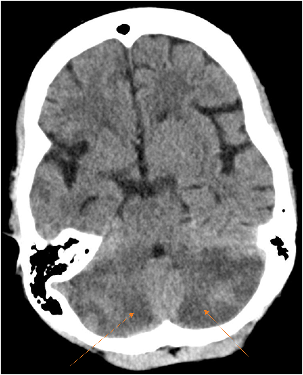
26A,B. Cerebellar infarct in the distribution of the posterior
Stroke in a Young Adult:Immunomodulation-Responsive Bilateral
The endovascular treatment of bilateral infarction of middle

Posterior inferior cerebellar artery

MRI finding in a patient with AICA artery territory infarction
Stroke Imaging: Practice Essentials, Computed Tomography, Magnetic

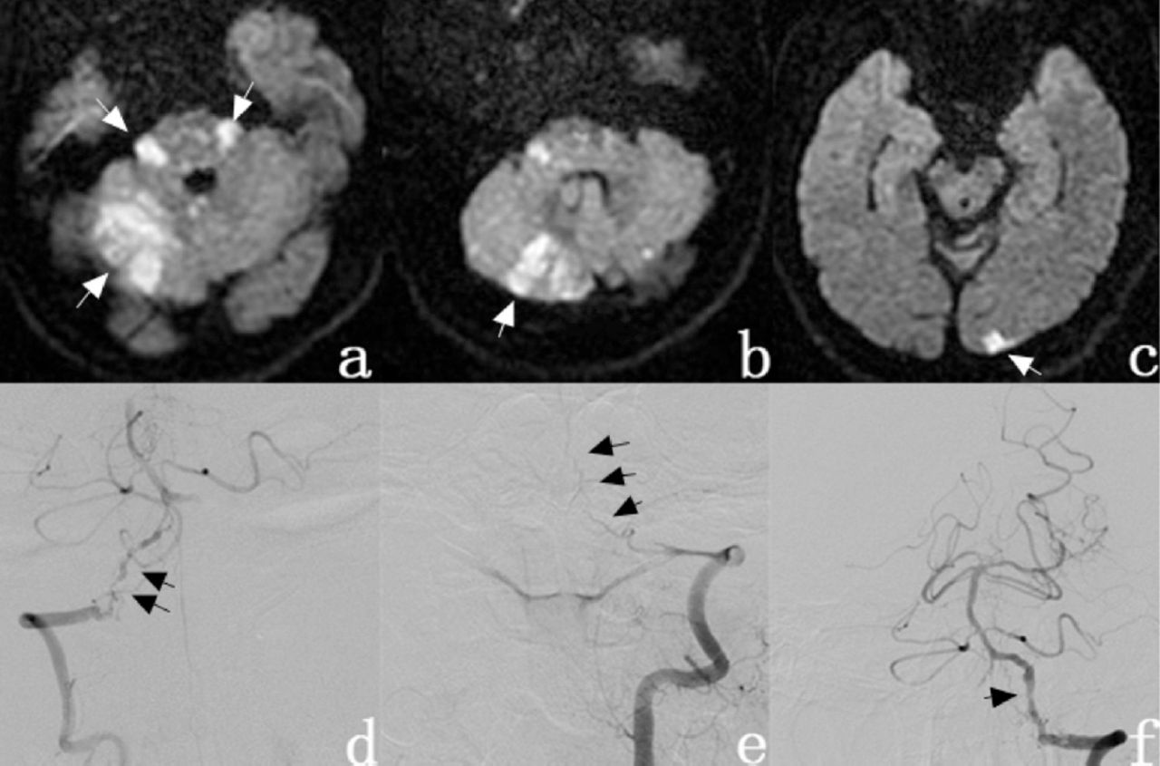
Cerebral angiogram which showed absence of the left Posterior

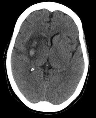
Posterior inferior cerebellar artery (PICA) infarct
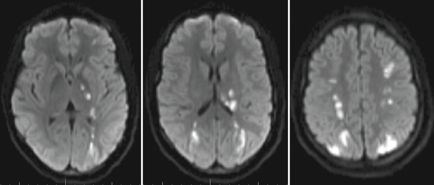
MRI on admission revealed multiple infarcts in the posterior

T2-weighted (A) and diffusion (B) magnetic resonance imaging